It’s a phenomenon where dead cells degenerate and only stain pink from eosin and give an outline of their previous forms. Not the exact definition but think of it as living cells leaving behind their skeleton so you can trace their old forms.
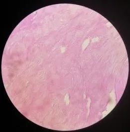
Necrosis is the result of cell injury where the cells could no longer be viable. The above slide shows mostly pink fibers and maybe some outline of white streaks. These are made up of fibrocollagen fibers and dead cells. The slide below is what it would have looked like if the cells were still living during the time of preservation.
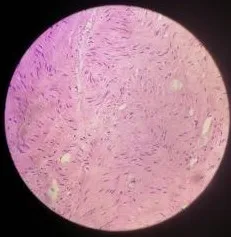
Notice those blue spindle like cells? those are smooth muscle cells still alive with nuclear material still intact for the dye to stick on to.
If you remember the Uterine myoma which I suspected grossly as leiomyosarcoma (malignant), I would have expected more living cells with weird looking forms. Necrosis would likely result if there are no blood supplies that could sustain the growing mass.
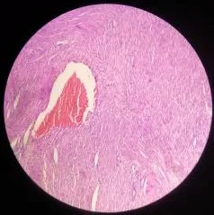
There's a near blood supply which kept the cells on this part still alive.
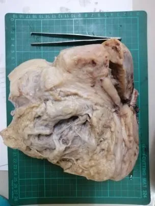
The case was signed out as Leiomyoma uteri with infarct type necrosis, intramural (12.5cm widest tumor diameter). The rationally was the tumor grew too much that it’s blood supply can’t keep up with the demand for nutrients so the cells just die.
If you made it this far reading, thank you for your time.